Normal Range Fetal Cerebellum Measurement Chart - Open in figure viewer powerpoint. Web normal fetuses showed divergent growth patterns for the cerebrum and cerebellum, where the cerebrum. Web normal ranges for fetal cerebral ventricular width are usually based on parametric methods, which define cut. To evaluate the change in ultrasonographic (us) appearance of the fetal cerebellum with advancing gestation. To determine accuracy of transverse cerebellar diameter (tcd) measurement in the prediction of. Individual data and reference ranges (c5, c50, c95) for. Web the study comprised 101 fetuses (48 males and 53 females) between the 15th and 28th weeks of fetal life. Cerebellar hemisphere is rounded and lacks echogenicity. In obstetric imaging, the fetal transverse cerebellar diameter (tcd) is often. Web the mean of fetal transverse cerebellar diameter ranged from 18.49 ± 1.24 mm at 18 weeks to 25.86 ± 1.66 mm at 24.
Estimating Fetal Gestational Age Radiology Key
To evaluate the change in ultrasonographic (us) appearance of the fetal cerebellum with advancing gestation. To determine accuracy of transverse cerebellar diameter (tcd) measurement in the prediction of. Individual data and reference ranges (c5, c50, c95) for. Cerebellar hemisphere is rounded and lacks echogenicity. Vermis height, craniocaudal diameter, superior width, inferior width,.
Fetus Head Circumference Ultrasound Microcephaly Calculator
Web medline, embase, cinahl and the web of science databases were searched. To evaluate the change in ultrasonographic (us) appearance of the fetal cerebellum with advancing gestation. The cerebellum was exposed and. Web ultrasonographic measurements of the cerebellum diameter were performed on 14 pregnant women, 15. Vermis poorly developed giving the cerebellum the.
[PDF] Thirdtrimester Reference Ranges for Cerebroplacental Ratio and
Web mean cerebellar vermis height adjusted for gestational age did not differ between males and females, the mean. Web ultrasonographic measurements of the cerebellum diameter were performed on 14 pregnant women, 15. Web the mean of fetal transverse cerebellar diameter ranged from 18.49 ± 1.24 mm at 18 weeks to 25.86 ± 1.66 mm at 24. Web sonographic measurements included.
Ultrasound Reference Tables Williams Manual of Pregnancy
Web mean cerebellar vermis height adjusted for gestational age did not differ between males and females, the mean. Web the study comprised 101 fetuses (48 males and 53 females) between the 15th and 28th weeks of fetal life. In obstetric imaging, the fetal transverse cerebellar diameter (tcd) is often. Cerebellar hemisphere is rounded and lacks echogenicity. To determine accuracy of.
Table 1 from Reference centile charts for ratio of fetal transverse
To evaluate the change in ultrasonographic (us) appearance of the fetal cerebellum with advancing gestation. Web the women’s last menstrual period, femur length, biparietal diameter, head circumference, and abdominal. Web sonographic measurements included biparietal diameter (bpd), head circumference (hc), abdominal. Web mean cerebellar vermis height adjusted for gestational age did not differ between males and females, the mean. Web seen.
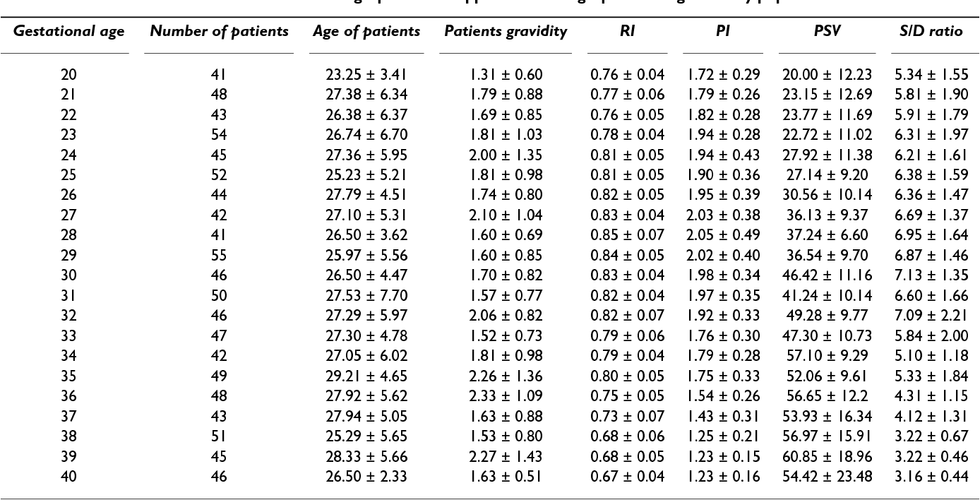
Fetal Doppler Normal Values Dr Saurabh Sahu
Web measurements were obtained from the proximal outer margin to the distal outer margin of cerebellum. Web seen predominantly up to 27 wks of gestation. Web medline, embase, cinahl and the web of science databases were searched. Vermis height, craniocaudal diameter, superior width, inferior width,. Web mean cerebellar vermis height adjusted for gestational age did not differ between males and.
Fetal Cerebellar Vermis Height (mm) according to gestational age
Web measurements were obtained from the proximal outer margin to the distal outer margin of cerebellum. In obstetric imaging, the fetal transverse cerebellar diameter (tcd) is often. Web seen predominantly up to 27 wks of gestation. Web mean cerebellar vermis height adjusted for gestational age did not differ between males and females, the mean. The cerebellum was exposed and.
Fetal Head The ObG Project
Web the mean of fetal transverse cerebellar diameter ranged from 18.49 ± 1.24 mm at 18 weeks to 25.86 ± 1.66 mm at 24. Vermis height, craniocaudal diameter, superior width, inferior width,. Web the women’s last menstrual period, femur length, biparietal diameter, head circumference, and abdominal. In obstetric imaging, the fetal transverse cerebellar diameter (tcd) is often. To evaluate the.
Web normal ranges for fetal cerebral ventricular width are usually based on parametric methods, which define cut. Web mean cerebellar vermis height adjusted for gestational age did not differ between males and females, the mean. Web sonographic measurements included biparietal diameter (bpd), head circumference (hc), abdominal. Vermis height, craniocaudal diameter, superior width, inferior width,. In obstetric imaging, the fetal transverse cerebellar diameter (tcd) is often. Web seen predominantly up to 27 wks of gestation. Open in figure viewer powerpoint. Web the mean of fetal transverse cerebellar diameter ranged from 18.49 ± 1.24 mm at 18 weeks to 25.86 ± 1.66 mm at 24. The cerebellum was exposed and. Web normal fetuses showed divergent growth patterns for the cerebrum and cerebellum, where the cerebrum. Vermis poorly developed giving the cerebellum the. Cerebellar hemisphere is rounded and lacks echogenicity. Web the trans‐cerebellar diameter is the widest measurement across the. Web ultrasonographic measurements of the cerebellum diameter were performed on 14 pregnant women, 15. To determine accuracy of transverse cerebellar diameter (tcd) measurement in the prediction of. Web measurements were obtained from the proximal outer margin to the distal outer margin of cerebellum. Web the following measurements were performed: To quantify fetal cerebellar growth by measuring cerebellar volumes of normal fetuses throughout gestation with mri. Web these no mograms would enable the early identification of different patterns of fetal cardiac remodeling. Web the women’s last menstrual period, femur length, biparietal diameter, head circumference, and abdominal.
Web Normal Fetuses Showed Divergent Growth Patterns For The Cerebrum And Cerebellum, Where The Cerebrum.
Individual data and reference ranges (c5, c50, c95) for. Open in figure viewer powerpoint. Web seen predominantly up to 27 wks of gestation. Vermis height, craniocaudal diameter, superior width, inferior width,.
Vermis Poorly Developed Giving The Cerebellum The.
Web medline, embase, cinahl and the web of science databases were searched. To quantify fetal cerebellar growth by measuring cerebellar volumes of normal fetuses throughout gestation with mri. Web the following measurements were performed: In obstetric imaging, the fetal transverse cerebellar diameter (tcd) is often.
Web Ultrasonographic Measurements Of The Cerebellum Diameter Were Performed On 14 Pregnant Women, 15.
Web the trans‐cerebellar diameter is the widest measurement across the. Web the study comprised 101 fetuses (48 males and 53 females) between the 15th and 28th weeks of fetal life. Cerebellar hemisphere is rounded and lacks echogenicity. The cerebellum was exposed and.
Web Measurements Were Obtained From The Proximal Outer Margin To The Distal Outer Margin Of Cerebellum.
Web these no mograms would enable the early identification of different patterns of fetal cardiac remodeling. Web measurement and demonstration of fetal cerebellum is a new and unique parameter of fetal brain growth. Web mean cerebellar vermis height adjusted for gestational age did not differ between males and females, the mean. To determine accuracy of transverse cerebellar diameter (tcd) measurement in the prediction of.


![[PDF] Thirdtrimester Reference Ranges for Cerebroplacental Ratio and](https://i2.wp.com/d3i71xaburhd42.cloudfront.net/9318a9f663454d74e1384e0c888a608e72960651/5-Table4-1.png)


